Brain Tumor: Symptoms, Causes, Diagnosis & Treatment
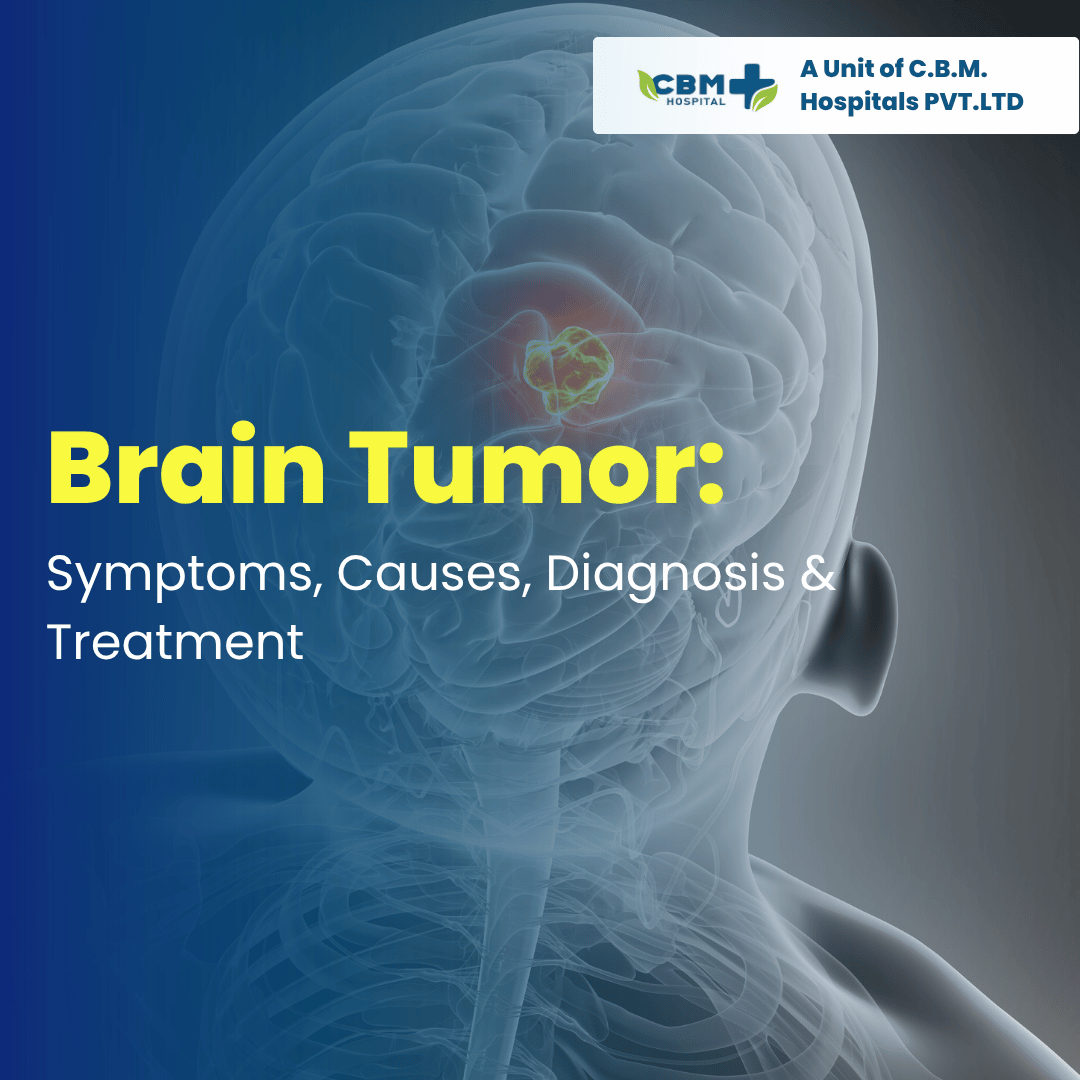
A brain tumor is an abnormal growth of cells in or around the brain. Brain tumors can be benign (non-cancerous) or malignant (cancerous). They can develop in different parts of the brain and may affect important functions such as movement, speech, memory, vision, balance, and coordination. Some brain tumors grow slowly, while others may grow more quickly. Symptoms depend on the tumor’s size, location, and type. Early medical evaluation is important when persistent or unusual neurological symptoms develop. This blog explains the symptoms, causes, types, diagnosis, treatment options, and recovery associated with brain tumors. What Is a Brain Tumor? A brain tumor develops when cells grow abnormally and form a mass within the brain or nearby structures. Brain tumors are broadly classified into: Benign Brain Tumors Benign tumors are generally non-cancerous and may grow slowly. However, they can still cause serious problems if they press on important areas of the brain or increase pressure inside the skull. Malignant Brain Tumors Malignant tumors are cancerous and may grow more aggressively. Some can invade nearby brain tissue or spread within the central nervous system. Brain tumors can also be classified as primary or secondary (metastatic). Primary brain tumors begin in the brain or nearby tissues. Secondary brain tumors occur when cancer from another part of the body spreads to the brain.   In many cases, the exact cause of a brain tumor is not known. Possible risk factors or associations may include: Certain genetic conditions Previous exposure to high-dose radiation to the head Family history of certain brain tumors Changes or mutations in specific genes A history of cancer elsewhere in the body, which can spread to the brain It is important to understand that having a risk factor does not necessarily mean that a person will develop a brain tumor. Types of Brain Tumors There are many types of brain tumors. Some commonly known types include: Gliomas Glioblastoma Meningioma Pituitary tumors Schwannomas Medulloblastomas Metastatic brain tumors The treatment depends greatly on the type, grade, location, and size of the tumor. Symptoms of a Brain Tumor Brain tumor symptoms vary from person to person. Some symptoms develop gradually, while others may appear more quickly. Common symptoms may include: Persistent or worsening headaches Nausea or vomiting Seizures Blurred or double vision Difficulty speaking Weakness or numbness Problems with balance or coordination Memory problems Changes in personality or behaviour Difficulty concentrating Excessive sleepiness Changes in hearing Difficulty walking Having one of these symptoms does not automatically mean that a person has a brain tumor. Many other medical conditions can cause similar symptoms. Headache and Brain Tumors Headaches are common and usually have many possible causes. A headache associated with a brain tumor may sometimes: Become progressively worse Be different from previous headaches Be accompanied by vomiting Occur along with neurological symptoms Be worse in the morning in some patients However, headache alone is usually not enough to diagnose a brain tumor. Seizures A seizure can sometimes be the first noticeable symptom of a brain tumor. A person may experience: Uncontrolled movements Temporary loss of awareness Staring episodes Abnormal sensations Confusion after the episode A first-time seizure requires medical evaluation. Warning Signs That Need Medical Attention Seek prompt medical attention if a person develops: A new or rapidly worsening headache A first-time seizure Sudden weakness or numbness New difficulty speaking Significant vision changes Increasing confusion Repeated unexplained vomiting Progressive problems with balance or walking Significant changes in behaviour or personality Sudden symptoms such as facial drooping, arm weakness, speech difficulty, or loss of consciousness can also indicate other medical emergencies, such as stroke, and require immediate emergency care. How Is a Brain Tumor Diagnosed? Doctors use several tests to determine whether a brain tumor is present and to understand its characteristics. Neurological Examination A doctor may evaluate: Muscle strength Reflexes Vision Eye movements Balance Coordination Memory Speech Sensation The examination helps identify which parts of the nervous system may be affected. MRI Scan An MRI of the brain is one of the most important imaging tests for evaluating suspected brain tumors. It can provide detailed information about: Tumor location Tumor size Surrounding brain tissue Possible swelling Relationship to important brain structures Contrast material may be used in some MRI examinations. CT Scan A CT scan can provide rapid images of the brain and may be particularly useful in emergency situations or when MRI is not suitable. Biopsy In some cases, a biopsy may be required to determine the exact type and grade of a tumor. A small sample of tissue is examined by a pathologist. The results can help doctors decide which treatment is most appropriate. Not every brain tumor requires a biopsy; the decision depends on the location and characteristics of the tumor. Brain Tumor Treatment Treatment depends on several factors, including: Type of tumor Grade Size Location Growth rate Symptoms Patient’s age and general health Whether the tumor has spread Treatment may involve one or more approaches. 1. Observation and Regular Monitoring Some slow-growing tumors that are not causing significant symptoms may be monitored with regular MRI scans and medical follow-up. This approach is sometimes called active surveillance or watchful waiting. 2. Brain Tumor Surgery Surgery may be recommended when the tumor can be safely removed or when tissue is needed for diagnosis. The main goals may include: Removing as much of the tumor as safely possible Reducing pressure on the brain Relieving symptoms Obtaining tissue for diagnosis The exact surgical approach depends on the tumor’s location and characteristics. Craniotomy A craniotomy involves temporarily removing a section of the skull to provide access to the brain. The surgeon removes or treats the tumor through this opening and then replaces the bone section. Minimally Invasive or Image-Guided Approaches For selected tumors, surgeons may use specialised techniques, navigation systems, or minimally invasive approaches to access the tumor while aiming to protect healthy brain tissue. Not every tumor is suitable for the same surgical technique. 3. Radiation Therapy Radiation therapy uses high-energy radiation to damage … Read more
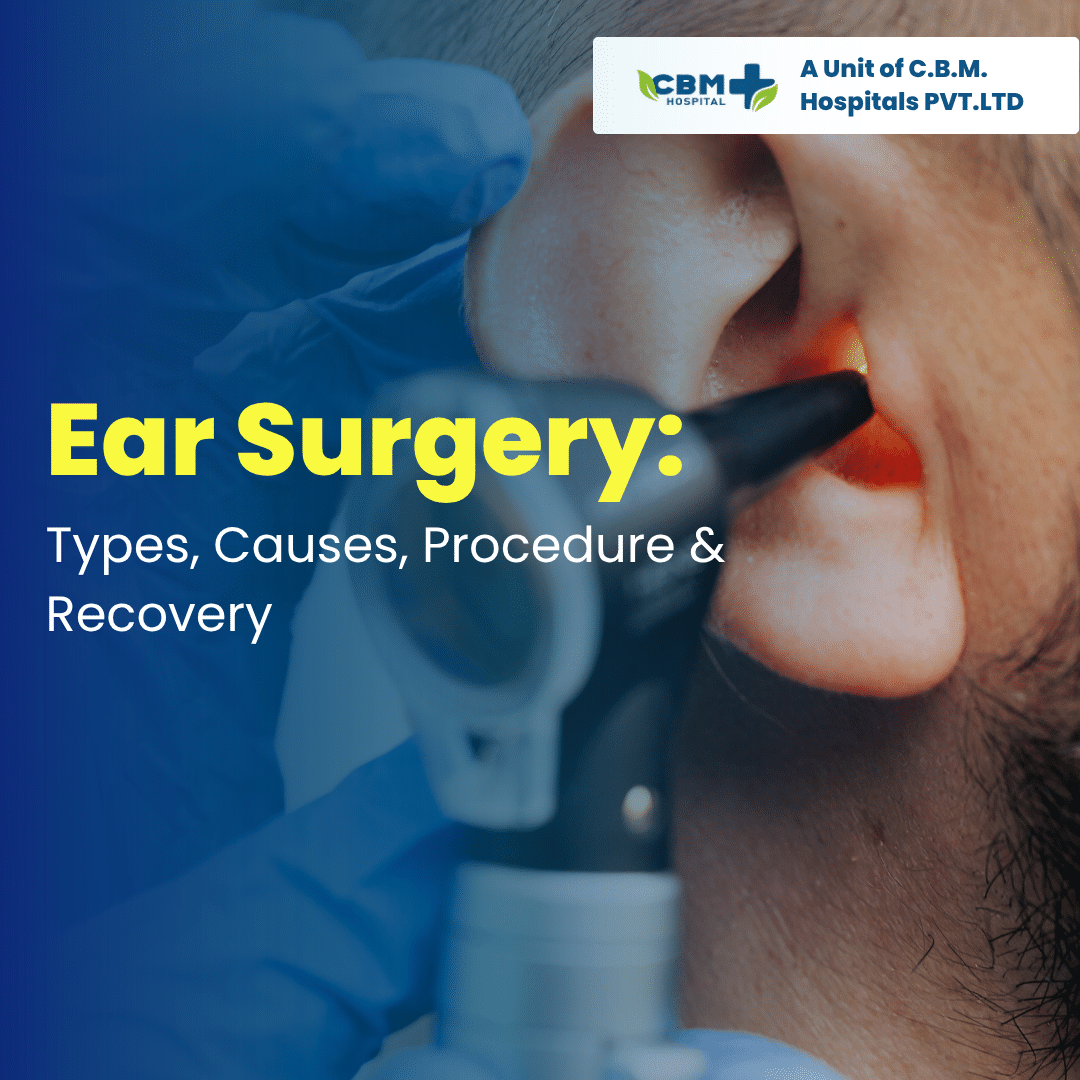
Ear Surgery: Types, Causes, Procedure & Recovery-

Ear problems can affect hearing, balance, ear health, and quality of life. Some ear conditions can be treated with medicines or simple procedures, while others may require surgery. Ear surgery, also known as otologic surgery, includes different procedures designed to treat problems affecting the outer, middle, or inner ear. Ear surgery may be recommended for conditions such as a damaged eardrum, chronic ear infection, hearing problems caused by middle-ear conditions, abnormal bone growth, or certain congenital problems. This blog explains the common types of ear surgery, reasons for surgery, procedure, benefits, risks, and recovery. What Is Ear Surgery? Ear surgery is a medical procedure performed to treat a problem affecting one or more parts of the ear. The ear has three main sections: Outer ear – includes the ear canal and visible ear. Middle ear – contains the eardrum and tiny hearing bones. Inner ear – contains structures involved in hearing and balance. The type of surgery depends on the location and cause of the ear problem. Why Is Ear Surgery Needed? Doctors may recommend ear surgery when a condition cannot be adequately treated with medicines or other non-surgical approaches. Common reasons include: Chronic or repeated ear infections Perforated or damaged eardrum Fluid buildup in the middle ear Hearing loss caused by middle-ear problems Cholesteatoma Abnormal growth of bone in the ear Damage to the small hearing bones Certain congenital ear abnormalities Some causes of dizziness or balance problems Certain tumors or other abnormal growths Not every ear problem requires surgery. An ENT specialist usually evaluates the symptoms and test results before recommending an operation. Common Types of Ear Surgery There are several types of ear surgery, and each is used for a specific condition. 1. Tympanoplasty Tympanoplasty is surgery to repair a perforated or damaged eardrum. A hole in the eardrum can occur because of: Ear infections Injury Sudden pressure changes Insertion of objects into the ear Previous ear surgery The surgeon repairs the damaged eardrum, sometimes using tissue taken from another part of the patient’s body. The goal is to close the perforation and, when possible, improve or protect hearing. 2. Myringoplasty Myringoplasty is a procedure specifically focused on repairing a perforation in the eardrum. It may be considered when the perforation does not heal naturally and causes problems such as repeated ear discharge or hearing difficulties. 3. Mastoidectomy A mastoidectomy involves removing diseased tissue from the mastoid bone located behind the ear. It may be performed for conditions such as: Chronic ear infection Cholesteatoma Infection involving the mastoid bone The exact surgical approach depends on the disease and its extent. 4. Stapedectomy or Stapedotomy These procedures are used to treat certain cases of otosclerosis, a condition in which abnormal bone growth affects movement of the stapes, one of the tiny bones involved in hearing. The surgeon modifies or replaces part of the affected structure to improve the transmission of sound. 5. Ossiculoplasty Ossiculoplasty is performed to repair or reconstruct the tiny bones of the middle ear when they have been damaged. These bones are called the: Malleus Incus Stapes Damage may occur because of chronic infection, trauma, or other ear conditions. 6. Grommet or Ear Tube Surgery Ear tubes, also called ventilation tubes or grommets, may be inserted into the eardrum to help drain fluid and improve ventilation of the middle ear. They are commonly considered for selected children with repeated ear infections or persistent middle-ear fluid, although they may also be used in some adults. 7. Cochlear Implant Surgery A cochlear implant may be considered for certain people with severe to profound hearing loss who receive limited benefit from conventional hearing aids. The implant uses an external component and an internal device to provide electrical stimulation to the auditory nerve. Cochlear implant treatment involves detailed hearing assessment and specialist evaluation. Symptoms That May Require an ENT Evaluation Not every symptom means surgery is required, but persistent symptoms should be assessed by an ENT specialist. Common symptoms include: Reduced hearing Repeated ear infections Persistent ear discharge Ear pain Ringing or buzzing in the ear Feeling of fullness or pressure Dizziness or balance problems Repeated fluid buildup A perforated eardrum Hearing difficulty affecting daily activities Warning Signs That Need Prompt Medical Attention Seek medical attention promptly if you experience: Sudden hearing loss Severe ear pain Significant swelling around the ear Blood or pus coming from the ear Severe dizziness Facial weakness High fever with ear symptoms Severe headache associated with an ear infection Sudden hearing loss, particularly when it develops rapidly, should be evaluated urgently. How Is an Ear Problem Diagnosed? Before recommending surgery, an ENT specialist may perform several examinations and tests. Ear Examination The doctor may use an otoscope or other specialised equipment to examine the ear canal and eardrum. Hearing Test An audiometry test can measure hearing ability and help determine the type and degree of hearing loss. Tympanometry Tympanometry evaluates the movement of the eardrum and can help identify problems involving the middle ear. Imaging A CT scan or MRI may be recommended for selected conditions, particularly when the doctor needs more detailed information about the ear and surrounding structures. What Happens During Ear Surgery? The exact procedure depends on the type of surgery. Generally, the process may involve: Medical and ENT evaluation before surgery. Hearing tests and imaging when required. Administration of local or general anaesthesia depending on the procedure. Accessing the affected part of the ear. Repairing, removing, or reconstructing the damaged structure. Closing the surgical area. Monitoring the patient after surgery. Some ear procedures are performed through the ear canal, while others may require an incision around or behind the ear. Is Ear Surgery Painful? Anaesthesia is used during surgery to prevent pain during the procedure. After surgery, some patients may experience: Mild to moderate discomfort Ear fullness Temporary dizziness Swelling Tenderness around the surgical area Pain medicines prescribed by the doctor can usually help manage post-operative discomfort. Benefits of Ear Surgery The potential benefits depend on the underlying condition … Read more

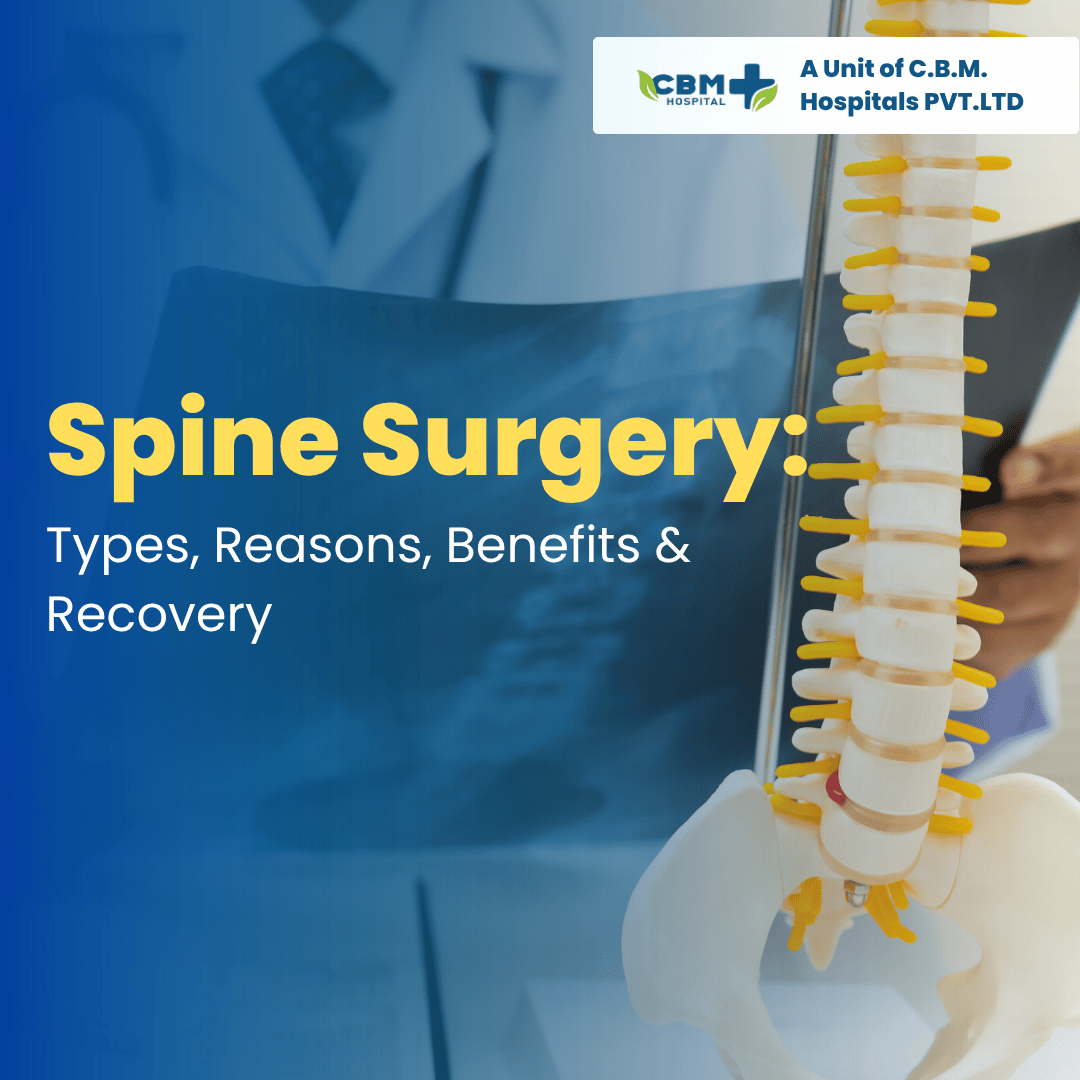
Spine Surgery: Types, Reasons, Benefits & Recovery-

Spine problems can affect people of different ages and may cause back pain, neck pain, stiffness, numbness, weakness, or difficulty walking. While many spinal conditions improve with medicines, physiotherapy, exercise, and lifestyle changes, some patients may need surgery when symptoms are severe or when the spine or nerves are under significant pressure. Spine surgery is performed to treat specific problems affecting the bones, discs, nerves, or spinal cord. The type of surgery depends on the patient’s condition, symptoms, imaging results, and overall health. This blog explains the common types of spine surgery, why surgery may be needed, its potential benefits, and what patients can expect during recovery. What Is Spine Surgery? Spine surgery refers to surgical procedures used to treat conditions affecting the spine, spinal discs, nerves, or spinal cord. The spine consists of vertebrae, discs, joints, ligaments, and nerves. Problems in any of these structures can sometimes cause pain or neurological symptoms. The main goals of spine surgery may include: Relieving pressure on nerves or the spinal cord Removing damaged or problematic tissue Stabilising an unstable section of the spine Correcting certain spinal deformities Treating fractures or serious injuries Improving movement and function Not every patient with back or neck pain requires surgery. Why Is Spine Surgery Needed? Doctors may recommend spine surgery when a spinal condition causes persistent or severe symptoms and non-surgical treatments have not provided sufficient improvement. Common reasons include: Herniated or slipped disc Spinal stenosis Spinal fractures Spinal instability Spondylolisthesis Certain spinal deformities Spinal cord or nerve compression Some spinal infections Certain spinal tumors Severe degenerative spinal conditions In some situations, surgery may be recommended sooner, especially when there is significant nerve or spinal cord compression. Common Symptoms That May Require Evaluation Spinal conditions can cause different symptoms depending on the affected area. Back and Neck Symptoms Persistent back pain Neck pain Stiffness Pain that travels into the arms or legs Difficulty bending or moving Muscle spasms Nerve-Related Symptoms Numbness or tingling Weakness in the arms or legs Burning or shooting pain Difficulty walking Loss of coordination Severe or rapidly worsening weakness, loss of bladder or bowel control, or numbness around the groin area can be an emergency and requires urgent medical attention. Types of Spine Surgery There are several types of spine surgery. The appropriate procedure depends on the patient’s diagnosis. 1. Discectomy A discectomy involves removing part or all of a damaged disc that is pressing on a spinal nerve. It may be considered for some patients with a herniated or slipped disc when nerve compression causes persistent pain, weakness, or other symptoms despite appropriate non-surgical treatment. Microdiscectomy A microdiscectomy uses a smaller surgical approach and specialised instruments to remove the portion of the disc pressing on the nerve. It is commonly used for certain lumbar disc problems. 2. Spinal Fusion Surgery Spinal fusion is performed to join two or more vertebrae so that they become more stable. The surgeon may use: Bone graft material Screws Rods Plates Other implants Fusion may be considered for conditions such as spinal instability, certain fractures, deformities, or selected degenerative conditions. 3. Laminectomy A laminectomy involves removing part of the vertebral bone called the lamina. The goal is usually to create more space for the spinal cord or nerves. It may be used in some patients with spinal stenosis, where narrowing of the spinal canal places pressure on nerves. 4. Laminoplasty Laminoplasty is another procedure used to create more space around the spinal cord, particularly in selected cases of cervical spinal stenosis. Instead of completely removing the lamina, the surgeon reshapes or repositions it to increase the available space. 5. Spinal Decompression Surgery Spinal decompression refers to procedures designed to relieve pressure on spinal nerves or the spinal cord. Depending on the condition, decompression may involve removing: Part of a disc Bone Thickened ligament Other tissue causing compression 6. Minimally Invasive Spine Surgery Some spinal procedures can be performed using minimally invasive techniques. These approaches generally use smaller incisions and specialised instruments. Depending on the procedure, potential advantages may include less disruption to surrounding tissues and a shorter recovery period. However, minimally invasive surgery is not appropriate for every spinal condition. When Is Spine Surgery Recommended? Spine surgery is not usually the first treatment for common back pain. Doctors may initially recommend: Medicines Physiotherapy Exercise Activity modification Weight management when appropriate Other non-surgical treatments Surgery may be considered when: Pain continues despite appropriate conservative treatment. A nerve is significantly compressed. There is progressive weakness or neurological deterioration. The spine is unstable. There is a serious fracture or injury. There is a spinal deformity requiring correction. A tumor or infection requires surgical treatment. Emergency compression of nerves or the spinal cord is present. The decision should be based on symptoms, physical examination, imaging, and the patient’s overall condition. How Is Spine Surgery Diagnosed and Planned? Before surgery, the doctor may perform a detailed neurological and physical examination. Tests may include: X-Ray X-rays can show the alignment and structure of the spine and may help identify fractures or certain degenerative changes. MRI Scan MRI provides detailed images of the discs, nerves, spinal cord, and surrounding soft tissues. CT Scan CT scans provide detailed images of the bones and may be particularly useful for evaluating fractures and certain spinal abnormalities. Nerve Tests In selected cases, tests such as nerve conduction studies or electromyography may be recommended to evaluate nerve function. Benefits of Spine Surgery The potential benefits depend on the underlying condition and the type of procedure. Possible benefits include: Relief from nerve-related pain Reduced pressure on nerves or the spinal cord Improved mobility Improved strength in selected patients Better ability to perform daily activities Stabilisation of an unstable spine Correction of certain spinal problems However, surgery does not guarantee complete pain relief, and outcomes vary from patient to patient. Risks of Spine Surgery Like any major procedure, spine surgery can have risks. Possible complications include: Infection Bleeding Blood clots Nerve injury Spinal fluid leakage Persistent or recurrent pain … Read more

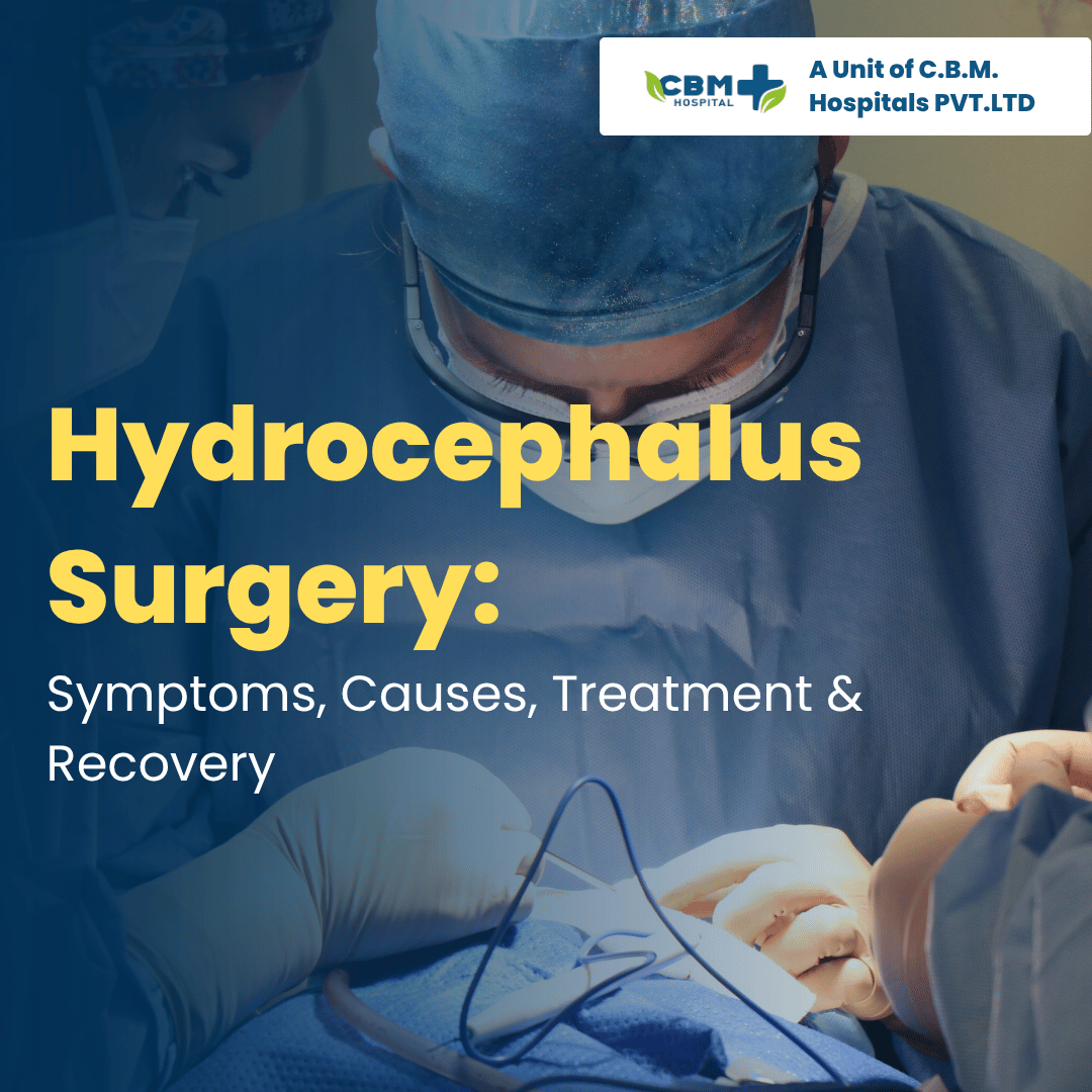
Hydrocephalus Surgery: Symptoms, Causes, Treatment & Recovery

Hydrocephalus is a condition in which excess cerebrospinal fluid (CSF) builds up inside the brain. This can increase pressure on the brain and may cause headaches, vomiting, difficulty walking, memory problems, vision changes, or other neurological symptoms. Hydrocephalus can affect babies, children, adults, and older people. Treatment depends on the cause, severity, age, and overall health of the patient. In many cases, hydrocephalus surgery is required to help drain the excess fluid and reduce pressure on the brain. This blog explains the symptoms, causes, diagnosis, treatment options, surgery, and recovery from hydrocephalus. What Is Hydrocephalus? Hydrocephalus occurs when CSF accumulates in the spaces (ventricles) inside the brain. Normally, CSF flows around the brain and spinal cord and is then absorbed into the bloodstream. When this flow or absorption is disrupted, fluid can build up. This may cause the ventricles to enlarge and increase pressure inside the skull. Hydrocephalus can develop suddenly or gradually and requires medical evaluation because untreated severe hydrocephalus can cause serious complications. What Causes Hydrocephalus? Hydrocephalus can have several causes, including: Blockage of normal CSF flow Brain tumors or other growths Brain infections such as meningitis Bleeding in or around the brain Head injury Congenital abnormalities present from birth Certain developmental conditions Problems with CSF absorption Age-related changes in some older adults Sometimes, the exact cause may not be immediately clear. Symptoms of Hydrocephalus Symptoms can vary depending on the patient’s age and how quickly fluid builds up. Symptoms in Adults and Older Adults Common symptoms may include: Persistent or worsening headache Nausea or vomiting Difficulty walking or balance problems Memory problems Confusion Difficulty concentrating Vision problems Excessive sleepiness Urinary urgency or loss of bladder control Changes in personality or behaviour In some older adults with normal pressure hydrocephalus (NPH), a combination of walking difficulty, memory changes, and urinary problems can occur. Symptoms in Babies and Young Children Signs may include: Rapid increase in head size Bulging soft spot on the baby’s head Poor feeding Vomiting Irritability Excessive sleepiness Developmental delays Seizures in some cases Eyes appearing directed downward Parents should seek medical attention if they notice unusual or progressive symptoms. When Is Hydrocephalus an Emergency? Sudden or severe symptoms may indicate increased pressure inside the brain and require urgent medical assessment. Seek emergency medical care if a person develops: Severe or rapidly worsening headache Repeated vomiting Loss of consciousness Seizures Severe confusion Sudden vision changes Difficulty speaking or moving Significant changes in alertness Hydrocephalus should not be self-diagnosed because similar symptoms can occur with other neurological conditions. How Is Hydrocephalus Diagnosed? A doctor may use a combination of neurological examination, medical history, and imaging tests. CT Scan A CT scan can show enlarged brain ventricles and may help identify bleeding, tumors, or other causes of fluid buildup. MRI Scan MRI provides detailed images of the brain and can help doctors understand the CSF pathways and identify possible blockages or other abnormalities. Neurological Examination Doctors may assess: Walking and balance Memory Eye movements Muscle strength Reflexes Coordination Additional tests may be recommended depending on the suspected type of hydrocephalus. Does Hydrocephalus Always Require Surgery? Not every patient is treated in exactly the same way. Treatment depends on the type and cause of hydrocephalus. Some patients may require close monitoring and treatment of the underlying condition. However, when excess CSF is causing significant pressure or symptoms, a procedure to improve fluid drainage may be necessary. The neurosurgeon decides the most appropriate treatment after evaluating the patient’s symptoms and brain imaging. Hydrocephalus Surgery The main goal of hydrocephalus surgery is to allow excess CSF to drain properly and reduce pressure on the brain. Two commonly used surgical approaches are shunt surgery and endoscopic third ventriculostomy (ETV). 1. Ventriculoperitoneal (VP) Shunt Surgery A VP shunt is one of the most commonly used treatments for hydrocephalus. During the procedure, a small tube is placed into one of the brain’s ventricles. The tube is connected to a valve and another catheter, which carries excess CSF to the abdomen. The fluid can then be absorbed naturally by the body. How Does a VP Shunt Work? The system generally consists of: A catheter placed inside the brain A valve that regulates CSF flow A second tube that carries fluid to the abdominal cavity The valve helps control the amount of fluid drained. 2. Endoscopic Third Ventriculostomy (ETV) ETV is another surgical option for selected patients. In this minimally invasive procedure, the neurosurgeon uses an endoscope to create a small opening in the floor of the third ventricle. This allows CSF to bypass a blockage and flow through another pathway. ETV may be appropriate for certain types of obstructive hydrocephalus, but it is not suitable for every patient. The choice between ETV and shunt surgery depends on factors such as the cause of hydrocephalus, age, imaging findings, and previous treatments. What Happens During Hydrocephalus Surgery? The exact procedure varies according to the treatment selected. Generally, the process may include: Pre-operative examination and brain imaging Administration of anesthesia Placement of the shunt or performance of the endoscopic procedure Testing the system or confirming appropriate CSF flow Closing the surgical openings Monitoring the patient after surgery The medical team will explain the specific procedure and possible risks before surgery. Risks of Hydrocephalus Surgery Like any surgical procedure, hydrocephalus surgery has potential risks. These may include: Infection Bleeding Shunt blockage Shunt malfunction Over-drainage or under-drainage of CSF Seizures Need for additional surgery Anesthesia-related complications A shunt may require monitoring throughout a patient’s life, and some patients may eventually need revision surgery if the system stops functioning properly. Recovery After Hydrocephalus Surgery Recovery depends on the patient’s age, type of hydrocephalus, cause, surgical procedure, and overall health. After surgery, doctors may monitor: Consciousness and neurological function Headache and vomiting Vision Walking and balance Signs of infection Shunt function, when applicable Some patients may return home after a short hospital stay, while others may need longer observation or rehabilitation. Recovery at Home Patients and caregivers should follow the neurosurgeon’s … Read more

Hernia: Symptoms, Types, Treatment and When Surgery Is Needed-

A hernia is a common medical condition that occurs when an internal organ or tissue pushes through a weak area in the surrounding muscle or connective tissue. Hernias most commonly develop in the abdomen or groin and may appear as a visible or noticeable bulge. Some hernias cause only mild discomfort, while others can become painful or lead to serious complications if the blood supply to the trapped tissue becomes blocked. Understanding the symptoms, types, causes, risk factors, treatment options, and when hernia surgery may be needed can help you recognize the condition and seek timely medical advice. What Is a Hernia? A hernia develops when tissue from inside the body pushes through a weakened area of the surrounding muscle or abdominal wall. A hernia may appear as a lump or bulge that becomes more noticeable when standing, coughing, lifting something heavy, or straining. In some cases, the bulge may disappear when lying down. Hernias can occur in different parts of the body, but they are particularly common in the abdominal wall and groin. A hernia generally does not disappear permanently without treatment. The appropriate management depends on the type of hernia, its size, symptoms, and risk of complications. What Causes a Hernia? A hernia can develop because of a combination of muscle weakness and increased pressure inside the abdomen. Factors that may contribute to a hernia include: Weak abdominal muscles Heavy lifting Chronic coughing Frequent straining during bowel movements Obesity Pregnancy Previous abdominal surgery Fluid accumulation in the abdomen Repeated physical strain Congenital weakness in the abdominal wall Increasing age Sometimes, a hernia develops gradually without one specific event causing it. What Are the Symptoms of a Hernia? The most common sign of a hernia is a visible or noticeable bulge. Other symptoms may include: A lump in the abdomen or groin Pain or discomfort at the site of the hernia A feeling of pressure or heaviness Burning or aching around the bulge Pain that becomes worse when coughing or lifting Discomfort while standing for long periods Difficulty with certain physical activities Some hernias may cause very few symptoms and are discovered during a routine examination. What Are the Different Types of Hernia? Hernias can occur in different locations. The most common types include: Inguinal Hernia An inguinal hernia occurs in the groin area and is the most common type of abdominal wall hernia. It can cause a bulge in the groin that may become more noticeable when coughing, standing, or lifting. Femoral Hernia A femoral hernia develops near the upper thigh or groin. It is less common than an inguinal hernia but may have a higher risk of becoming trapped. Umbilical Hernia An umbilical hernia develops around the belly button. It can occur in children and adults. In adults, factors such as obesity, pregnancy, or increased abdominal pressure may contribute to its development. Incisional Hernia An incisional hernia can develop through or near a previous surgical incision in the abdominal wall. It may occur months or even years after abdominal surgery. Hiatal Hernia A hiatal hernia occurs when part of the stomach moves upward through an opening in the diaphragm. Unlike abdominal wall hernias, a hiatal hernia may not cause an external bulge. It can sometimes contribute to symptoms such as acid reflux and heartburn. What Makes a Hernia Worse? Certain activities and conditions can increase pressure inside the abdomen and make hernia symptoms more noticeable. These may include: Heavy lifting Persistent coughing Sneezing Straining during bowel movements Obesity Certain strenuous activities Prolonged standing If you have a known hernia, discuss appropriate activity levels with your doctor rather than completely avoiding physical activity without guidance. Warning Signs of a Hernia Complication Most hernias are not immediately life-threatening, but a hernia can sometimes become incarcerated or strangulated. An incarcerated hernia becomes trapped and cannot be pushed back into the abdomen. A strangulated hernia occurs when the blood supply to the trapped tissue becomes compromised. Seek urgent medical attention if you develop: Sudden or severe pain at the hernia site A bulge that becomes hard or very tender A bulge that cannot be pushed back when it previously could Red, purple, or dark discoloration over the hernia Nausea or repeated vomiting Abdominal swelling Difficulty passing stool or gas Fever along with severe abdominal symptoms A strangulated hernia can damage the affected tissue and may require emergency surgery. How Is a Hernia Diagnosed? Doctors usually diagnose a hernia through a physical examination. The doctor may ask you to stand, cough, or strain gently while examining the affected area. The examination can help identify: The location of the hernia The size of the bulge Whether it is tender Whether it can be pushed back Whether there are signs of complications Ultrasound An ultrasound may be recommended when the diagnosis is unclear or when the hernia is difficult to detect during examination. CT Scan A CT scan can provide detailed images of the abdominal wall and internal organs. It may be useful when complications are suspected or when the diagnosis is uncertain. The appropriate investigation depends on the type of suspected hernia and the patient’s symptoms. Does Every Hernia Require Surgery? No. Treatment depends on the type and symptoms of the hernia. Some small hernias that cause little or no discomfort may be monitored by a doctor. However, many symptomatic hernias eventually require surgical repair because the underlying weakness in the abdominal wall generally does not repair itself on its own. Your doctor will consider factors such as: Hernia size Location Symptoms Changes in the size of the hernia Overall health Risk of complications Effect on daily activities When Is Hernia Surgery Needed? Hernia surgery may be recommended when: The hernia causes persistent or significant pain The hernia is increasing in size Symptoms interfere with daily activities The hernia becomes difficult to push back There is a risk of complications The hernia becomes incarcerated or strangulated Emergency surgery may be necessary if the blood supply to trapped tissue is compromised. How Is … Read more

Appendicitis: Symptoms, Causes and When Surgery Is Needed

Appendicitis is a medical condition in which the appendix becomes inflamed. The appendix is a small, tube-shaped structure attached to the large intestine, usually located in the lower-right side of the abdomen. Appendicitis can develop suddenly and may become serious if it is not treated promptly. In some cases, the inflamed appendix can burst, allowing infection to spread inside the abdomen. Understanding the symptoms, causes, risk factors, diagnosis, treatment options, and when appendicitis surgery may be needed can help you recognize the condition and seek timely medical care. What Is Appendicitis? Appendicitis occurs when the appendix becomes inflamed and swollen. The appendix does not have a major role in digestion, but inflammation can cause significant abdominal pain and other symptoms. Appendicitis can affect people of all ages, although it is more common in children, teenagers, and young adults. Because symptoms can sometimes resemble other abdominal conditions, medical evaluation is important when appendicitis is suspected. What Causes Appendicitis? Appendicitis usually develops when the opening of the appendix becomes blocked. The blockage can allow bacteria to multiply and cause inflammation and swelling. Possible causes of blockage include: Hardened stool Swelling of lymphoid tissue Intestinal infections Inflammatory bowel conditions Parasites in rare cases Other blockages In some patients, the exact cause cannot be identified. Once inflammation begins, pressure inside the appendix may increase. Without appropriate treatment, the appendix can become severely damaged and may eventually rupture. What Are the Symptoms of Appendicitis? The symptoms of appendicitis can vary, but abdominal pain is one of the most common signs. Typical symptoms include: Pain that may begin around the belly button Pain that moves toward the lower-right abdomen Increasing abdominal pain Loss of appetite Nausea Vomiting Mild fever Abdominal tenderness Bloating Constipation or diarrhea in some cases Appendicitis pain may become worse when walking, coughing, sneezing, or making sudden movements. Not everyone experiences all of these symptoms, and the location of pain may be different in some people, particularly pregnant women. How Does Appendicitis Pain Usually Start? Appendicitis pain often begins near the belly button and may later move to the lower-right side of the abdomen. The pain may initially be mild but can become stronger and more constant over time. However, abdominal pain can have many different causes. Therefore, the pattern of pain alone cannot confirm appendicitis. If abdominal pain is severe, persistent, or worsening, medical evaluation is important. Warning Signs That Need Urgent Medical Attention Appendicitis can become dangerous if the appendix ruptures. Seek urgent medical attention if you experience: Severe or rapidly worsening abdominal pain Severe tenderness in the abdomen Persistent vomiting Fever or chills Increasing abdominal swelling Severe weakness or confusion Abdominal pain followed by sudden worsening or a temporary decrease in pain A ruptured appendix can cause peritonitis, a serious infection of the lining of the abdomen, and may require emergency treatment. Do not try to treat suspected appendicitis at home or wait for severe symptoms to develop. How Is Appendicitis Diagnosed? Doctors usually begin with a medical history and physical examination. The doctor may ask about: When the pain started Where the pain is located Whether the pain has moved Associated symptoms such as vomiting or fever Changes in bowel movements Previous medical conditions During the examination, the doctor may gently press different areas of the abdomen to check for tenderness and other signs of inflammation. Blood Tests Blood tests may help identify signs of infection or inflammation. A raised white blood cell count can occur with appendicitis, although blood tests alone cannot confirm the diagnosis. Urine Tests A urine test may be performed to help rule out urinary tract infections or kidney stones, which can sometimes cause similar symptoms. Ultrasound An abdominal ultrasound may be used to examine the appendix, particularly in children and pregnant patients. CT Scan A CT scan can provide detailed images of the abdomen and may help confirm appendicitis or identify complications such as an abscess or perforation. The choice of imaging depends on the patient’s age, symptoms, examination findings, and clinical situation. Can Appendicitis Be Treated Without Surgery? In selected patients, doctors may consider treatment with antibiotics, particularly in certain uncomplicated cases. However, antibiotics are not suitable for everyone, and appendicitis can recur after non-operative treatment. The appropriate treatment depends on factors such as: Whether the appendix has ruptured Severity of inflammation Presence of an abscess Overall health Imaging findings The surgeon’s assessment Patients should not take antibiotics without medical evaluation. When Is Appendicitis Surgery Needed? Appendectomy, or surgical removal of the appendix, is a common treatment for appendicitis. Surgery may be recommended when: Appendicitis is confirmed Symptoms are worsening There is a high risk of appendix rupture The appendix has already perforated An abscess or other complication has developed Non-surgical treatment is not appropriate or has failed The timing of surgery depends on the patient’s condition and whether complications are present. What Is Appendectomy? An appendectomy is a surgical procedure in which the inflamed appendix is removed. There are two main approaches: Laparoscopic Appendectomy Laparoscopic surgery is performed through several small incisions. A camera and specialized surgical instruments are inserted through these small openings. The surgeon identifies and removes the appendix. Potential advantages may include: Smaller incisions Less postoperative discomfort for some patients Shorter hospital stay in appropriate cases Faster return to normal activities compared with some open procedures Open Appendectomy An open appendectomy involves a larger incision in the abdomen to remove the appendix. It may be required in certain situations, particularly when the appendix has ruptured or there is extensive infection, although the approach depends on the individual case and surgeon’s assessment. What Happens After Appendicitis Surgery? After an appendectomy, patients are monitored for recovery and possible complications. Recovery may include: Pain management Wound care Gradual return to normal eating Walking as advised Prescribed medicines when required Follow-up with the surgical team If the appendix has ruptured or there is significant infection, recovery may take longer and additional treatment may be required. Patients should follow their surgeon’s instructions regarding … Read more

Angioplasty: When Is It Needed and How Is It Performed?

Angioplasty, also known as percutaneous coronary intervention (PCI), is a minimally invasive procedure used to improve blood flow through narrowed or blocked arteries. It is commonly performed to treat coronary artery disease, a condition in which the blood vessels supplying the heart become narrowed because of plaque buildup. Angioplasty may be planned for people with certain types of coronary artery disease or performed urgently during a heart attack to restore blood flow to the heart. Understanding what angioplasty is, when it is needed, how the procedure is performed, its benefits and risks, recovery, and prevention can help patients understand their treatment options. What Is Angioplasty? Angioplasty is a procedure that opens a narrowed or blocked artery so that blood can flow more easily. The procedure is usually performed using a thin, flexible tube called a catheter. The catheter is inserted through a blood vessel, commonly in the wrist or groin, and guided toward the affected coronary artery. A small balloon at the tip of the catheter may then be inflated at the narrowed area. This helps widen the artery. In many cases, a small mesh tube called a stent is placed inside the artery to help keep it open. Angioplasty does not remove all the underlying causes of coronary artery disease, so medicines and lifestyle changes remain important after the procedure. Why Do Coronary Arteries Become Blocked? The coronary arteries can become narrowed because of atherosclerosis. Atherosclerosis occurs when fatty deposits, cholesterol, calcium, and other substances build up inside the artery walls. These deposits are commonly called plaques. As plaque increases, the artery can become narrower and restrict blood flow to the heart. This may cause angina, which is chest discomfort that occurs when the heart muscle does not receive enough oxygen-rich blood. If a plaque ruptures and a blood clot suddenly blocks an artery, it can cause a heart attack. When Is Angioplasty Needed? Angioplasty may be recommended when a coronary artery is significantly narrowed or blocked and restoring blood flow is expected to provide benefit. It may be considered for: Certain heart attacks Severe or persistent angina Significant narrowing of a coronary artery Coronary artery disease that is causing troublesome symptoms Situations where medicines and lifestyle changes do not adequately control symptoms During an acute heart attack, emergency angioplasty may be used to quickly restore blood flow to the heart. Not everyone with a blocked or narrowed artery requires angioplasty. The decision depends on the location and severity of the blockage, symptoms, heart function, overall health, and other factors. What Are the Symptoms That May Lead to Angioplasty? People with coronary artery disease may experience: Chest pressure or tightness Chest pain during physical activity Shortness of breath Unusual tiredness Pain spreading to the arm, shoulder, neck, jaw, or back Reduced ability to exercise Some people may have coronary artery disease without obvious symptoms. If chest discomfort is sudden, severe, persistent, or associated with sweating, shortness of breath, nausea, or fainting, it should be treated as a potential emergency. How Is Angioplasty Performed? Before angioplasty, the patient undergoes medical evaluation. Doctors may perform tests such as an ECG, blood tests, echocardiogram, or coronary angiography, depending on the situation. The procedure generally involves the following steps: 1. Preparing the Patient The patient is positioned in the procedure room and connected to monitors that track heart rate, blood pressure, and oxygen levels. The access site, usually the wrist or groin, is cleaned and numbed with local anesthesia. 2. Inserting the Catheter A small tube called a sheath is placed into the blood vessel. The cardiologist guides a thin catheter through the blood vessel toward the coronary arteries. 3. Identifying the Blockage A contrast dye is injected into the coronary arteries. X-ray imaging, called angiography, allows the doctor to see the blood vessels and identify narrowed or blocked areas. 4. Opening the Narrowed Artery A thin guidewire is passed through the affected area. A small balloon is then positioned at the blockage and inflated for a short period. The balloon presses the plaque against the artery wall and widens the passage for blood flow. 5. Placing a Stent In many cases, a stent is placed in the artery. The stent expands with the balloon and remains inside the artery to help maintain its opening. Most coronary stents used today are drug-eluting stents, which release medication designed to reduce the risk of tissue growing excessively inside the stent. 6. Completing the Procedure The balloon and catheter are removed while the stent remains in place. The access site is then treated to prevent bleeding, and the patient is monitored during recovery. Is Angioplasty Painful? Angioplasty is generally performed under local anesthesia, so the patient is usually awake during the procedure. The patient may feel pressure at the catheter insertion site. Some people may also experience temporary chest discomfort when the balloon is inflated. The medical team monitors the patient throughout the procedure and can provide medication if necessary. How Long Does Angioplasty Take? The duration varies depending on the number and complexity of blockages being treated. A straightforward procedure may take around an hour, while complex or emergency procedures can take longer. After angioplasty, the patient is monitored to check for bleeding, heart rhythm changes, chest pain, and other potential complications. What Are the Benefits of Angioplasty? For appropriately selected patients, angioplasty can provide important benefits, including: Restoring blood flow through a narrowed artery Relieving angina symptoms Improving blood supply to the heart muscle Improving exercise tolerance in some patients Reducing heart muscle damage during certain heart attacks Improving quality of life In an appropriate emergency heart attack setting, rapid restoration of blood flow can be particularly important. What Are the Risks of Angioplasty? Angioplasty is commonly performed, but like any medical procedure, it carries potential risks. Possible complications include: Bleeding or bruising at the catheter insertion site Blood vessel damage Abnormal heart rhythms Allergic reaction to contrast dye Blood clots Re-narrowing of the artery Heart attack Stroke Kidney problems related to contrast … Read more

Heart Attack: Symptoms, Causes and Emergency Treatment

A heart attack, medically known as a myocardial infarction (MI), is a serious medical emergency that occurs when blood flow to part of the heart muscle becomes blocked. Without enough blood and oxygen, the affected heart muscle can become damaged. A heart attack can happen suddenly, and getting emergency medical treatment as quickly as possible can reduce heart damage and improve the chances of recovery. Understanding the symptoms, causes, risk factors, warning signs, diagnosis, emergency treatment, and prevention of heart attack can help you recognize this potentially life-threatening condition and seek immediate medical care. What Is a Heart Attack? The heart needs a continuous supply of oxygen-rich blood through the coronary arteries. A heart attack usually occurs when a coronary artery becomes blocked, most often because of a blood clot forming over a ruptured or damaged fatty plaque. When blood flow is reduced or completely blocked, the heart muscle supplied by that artery does not receive enough oxygen. If the blockage is not treated quickly, part of the heart muscle can become permanently damaged. A heart attack is different from cardiac arrest. During cardiac arrest, the heart suddenly stops pumping effectively. A heart attack can sometimes lead to cardiac arrest, but the two conditions are not the same. What Causes a Heart Attack? The most common underlying cause of a heart attack is coronary artery disease (CAD). Over time, cholesterol, fat, and other substances can build up inside the walls of coronary arteries. This process is called atherosclerosis. A plaque can sometimes rupture, causing a blood clot to form. If the clot blocks blood flow through a coronary artery, a heart attack can occur. Other, less common causes can include severe coronary artery spasm or spontaneous tearing of a coronary artery. Risk Factors for Heart Attack Several factors can increase the risk of heart disease and heart attack, including: High blood pressure High cholesterol Diabetes Smoking or tobacco use Obesity or excess body weight Physical inactivity Unhealthy diet Family history of heart disease Increasing age Excessive alcohol consumption Chronic stress Previous heart disease Having one or more risk factors does not mean that a person will definitely have a heart attack, but managing these factors can help reduce cardiovascular risk. What Are the Symptoms of a Heart Attack? Heart attack symptoms can vary from person to person. Some attacks begin suddenly, while others may start gradually. Common symptoms include: Pressure, tightness, squeezing, or pain in the chest Pain or discomfort spreading to the arm, shoulder, back, neck, jaw, or upper abdomen Shortness of breath Cold sweating Nausea or vomiting Dizziness or light-headedness Unusual weakness or tiredness A feeling of anxiety or impending danger Chest discomfort is one of the most important warning signs, but not every heart attack causes severe chest pain. Women, older adults, and people with diabetes may sometimes experience less typical symptoms, such as unusual fatigue, shortness of breath, nausea, or back and jaw discomfort. Warning Signs That Need Emergency Medical Attention A suspected heart attack should always be treated as a medical emergency. Call emergency medical services immediately if you or someone around you develops: Chest pressure or pain that lasts several minutes or keeps returning Chest discomfort with sweating or shortness of breath Pain spreading to the arm, jaw, neck, or back Sudden severe weakness or dizziness Fainting or loss of consciousness Severe breathing difficulty Do not wait for symptoms to disappear on their own. If a heart attack is suspected, getting emergency medical help quickly is more important than trying to travel to a hospital independently. What Should You Do During a Suspected Heart Attack? If someone develops symptoms that may indicate a heart attack: Call emergency medical services immediately. Keep the person seated or resting in a comfortable position. Avoid unnecessary physical activity. Follow the instructions provided by emergency medical professionals. If the person becomes unresponsive and is not breathing normally, begin CPR and use an AED if available, following emergency guidance. Do not delay emergency care while trying home remedies. How Is a Heart Attack Diagnosed? Doctors use several tests to determine whether a heart attack has occurred. Electrocardiogram (ECG) An ECG records the electrical activity of the heart. It can help doctors identify changes associated with a heart attack and determine the type of heart attack. Blood Tests Blood tests, particularly cardiac troponin, can help detect damage to heart muscle. Doctors may perform repeated blood tests because cardiac markers can change over time. Echocardiogram An echocardiogram uses ultrasound to create images of the heart. It can help assess heart function and identify areas that are not moving normally. Coronary Angiography Coronary angiography can show blockages or narrowing in the coronary arteries. It may be performed during emergency treatment when doctors need to identify and treat a blocked artery. Other investigations may be recommended depending on the patient’s condition. Emergency Treatment for a Heart Attack Treatment depends on the type of heart attack, the location and severity of the blockage, and how quickly the patient reaches medical care. The main goal is to restore blood flow to the heart as quickly as possible and prevent further heart muscle damage. Treatment may include medicines and procedures. Medicines Doctors may use medications to: Prevent or reduce blood clotting Improve blood flow Reduce the workload on the heart Control pain Manage blood pressure Treat abnormal heart rhythms The specific medicines used depend on the patient’s condition and should be given under medical supervision. Angioplasty and Stent Placement Percutaneous coronary intervention (PCI) is a common emergency treatment for certain heart attacks. During the procedure, a thin catheter is guided through a blood vessel to the blocked coronary artery. A balloon may be used to open the narrowed area, and a stent is commonly placed to help keep the artery open. Rapid restoration of blood flow can limit damage to the heart muscle. Thrombolytic Treatment In certain situations where timely PCI is not available, doctors may consider clot-dissolving medicines, also known as thrombolytics. Whether this treatment … Read more

Knee Replacement Surgery: When Is It Needed and How Does It Work? –

Knee replacement surgery is a procedure used to relieve severe knee pain and improve movement when the knee joint has become significantly damaged. It is commonly recommended for people with advanced arthritis or other conditions that cause long-term knee pain, stiffness, and difficulty walking or performing everyday activities. The knee joint plays an important role in standing, walking, climbing stairs, and many other movements. When the cartilage covering the bones becomes severely damaged, the bones may rub against each other, causing pain and stiffness. Understanding the symptoms, causes, treatment options, types of knee replacement, surgical procedure, recovery, and when surgery may be needed can help patients make informed decisions with their doctor. What Is Knee Replacement Surgery? Knee replacement surgery, also called knee arthroplasty, involves replacing damaged parts of the knee joint with artificial components. The knee is made up of three main areas: Femur — the thigh bone Tibia — the shin bone Patella — the kneecap During knee replacement, the damaged surfaces of the knee are removed and replaced with specially designed artificial components. The goal is to reduce pain, improve joint function, and help the patient return to daily activities. Knee replacement does not necessarily mean that the entire knee is replaced. Depending on the condition, a surgeon may recommend total knee replacement or partial knee replacement. Why Does a Knee Need Replacement? The most common reason for knee replacement is severe osteoarthritis. Osteoarthritis gradually damages the cartilage inside the joint, leading to pain and stiffness. Other conditions that may damage the knee include: Rheumatoid arthritis Previous serious knee injury Fractures involving the knee joint Severe cartilage damage Joint deformity Other degenerative knee conditions Age-related changes can increase the risk of knee problems, but severe knee arthritis can affect people at different ages. What Are the Symptoms of Severe Knee Damage? Knee problems can develop gradually. Common symptoms include: Persistent knee pain Knee stiffness Difficulty walking Pain while climbing stairs Difficulty getting up from a chair Swelling around the knee Reduced knee movement Knee pain that becomes worse with activity Difficulty performing daily activities Pain that may interfere with sleep Some people also experience a grinding or clicking sensation in the knee. However, not everyone with knee pain requires replacement surgery. A proper medical evaluation is necessary to determine the cause and severity of the problem. When Is Knee Replacement Surgery Needed? Knee replacement surgery is generally considered when severe knee damage causes persistent pain and significantly affects daily life, particularly when non-surgical treatments have not provided sufficient relief. A doctor may consider surgery when: Knee pain is persistent or severe Walking becomes difficult Daily activities are significantly affected Knee stiffness limits movement Pain interferes with sleep There is significant joint damage on imaging Medicines and physiotherapy provide inadequate relief Other non-surgical treatments have been tried without sufficient improvement The decision is based on symptoms, physical examination, imaging, overall health, and how much the knee problem affects quality of life. Do All Knee Pain Patients Need Knee Replacement? No. Knee pain can have many causes, and knee replacement is not the first treatment for most people. Depending on the cause, doctors may initially recommend: Weight management when appropriate Physiotherapy Muscle-strengthening exercises Activity modification Pain-relieving medicines Assistive devices when required Other non-surgical treatments Knee replacement is generally considered when the knee has significant structural damage and symptoms remain troublesome despite appropriate treatment. Types of Knee Replacement Surgery There are different types of knee replacement procedures depending on how much of the joint is damaged. Total Knee Replacement In total knee replacement, the damaged surfaces of the knee are replaced with artificial components. This is commonly performed when arthritis or other damage affects multiple parts of the knee. Partial Knee Replacement A partial knee replacement replaces only the damaged portion of the knee. It may be suitable for selected patients whose disease is limited to one compartment of the knee and who meet other clinical requirements. The surgeon determines which procedure is appropriate after evaluating the knee. How Does Knee Replacement Surgery Work? Before surgery, the patient undergoes a medical assessment. This may include a physical examination, blood tests, imaging, and other tests depending on overall health. During the operation: The patient receives appropriate anesthesia. The surgeon makes an incision around the knee. Damaged cartilage and small amounts of damaged bone are removed. The damaged surfaces are prepared for the artificial components. The replacement components are positioned and secured. The surgeon checks the movement and stability of the knee. The incision is closed. The exact surgical technique can vary depending on the patient’s condition and the type of knee replacement being performed. What Happens After Knee Replacement Surgery? After surgery, the healthcare team monitors the patient and begins the recovery process. Patients are usually encouraged to start moving the knee and walking with assistance as advised by the medical team. Recovery may involve: Physiotherapy Walking exercises Knee-strengthening exercises Pain management Wound care Gradual increase in daily activities Follow-up appointments Physiotherapy is particularly important because it helps restore knee movement and strengthen the muscles around the joint. How Long Does It Take to Recover From Knee Replacement? Recovery is different for every patient. Many people gradually return to normal daily activities over the weeks and months following surgery. However, complete recovery can take longer depending on factors such as age, general health, muscle strength, the condition of the knee before surgery, and adherence to rehabilitation. Patients should follow their surgeon’s and physiotherapist’s instructions regarding walking, exercises, driving, work, and other activities. What Are the Benefits of Knee Replacement Surgery? For appropriately selected patients, knee replacement can provide significant benefits. Potential benefits include: Reduction in chronic knee pain Improved ability to walk Better knee movement Improved ability to perform daily activities Improved quality of life Greater independence in routine activities The outcome varies between individuals, and knee replacement cannot guarantee that every patient will become completely pain-free. Are There Risks Associated With Knee Replacement? Like any major surgery, knee replacement … Read more

Slip Disc: Symptoms, Causes, Treatment & When Surgery Is Needed-

Slip Disc: Symptoms, Causes, Treatment & When Surgery Is Needed  A slip disc, also known as a herniated disc or prolapsed disc, is a common spinal condition that can cause back pain, neck pain, numbness, or weakness. It occurs when the soft inner portion of a spinal disc pushes through a weakened or damaged outer layer.    Slip disc can affect people of different ages and may occur in the lower back, neck, or, less commonly, the upper and middle back. While many cases improve with medicines, activity modification, and physiotherapy, some people may require further treatment or surgery.    Understanding the symptoms, causes, risk factors, diagnosis, treatment options, and warning signs of a slip disc can help you seek medical attention at the right time.    What Is a Slip Disc?  The spine is made up of multiple bones called vertebrae. Between most of these bones are flexible structures called intervertebral discs. These discs act as cushions and help the spine absorb shock during movement. Each disc has a tough outer layer and a softer inner portion. When the outer layer becomes weak or develops a tear, the inner material can bulge or move outward. This is called a herniated or slipped disc. A slipped disc can sometimes press against nearby spinal nerves. This nerve compression can cause pain, tingling, numbness, or weakness in the area supplied by the affected nerve. Slip disc most commonly affects the lower back, but it can also occur in the neck. What Causes a Slip Disc?  A slip disc usually develops because of gradual wear and tear of the spinal discs. As people age, discs can become less flexible and more vulnerable to injury. Common causes and risk factors include: Age-related changes in the spine Incorrect lifting of heavy objects Sudden twisting or bending movements Poor posture Repetitive physical activities Being overweight or obese Lack of regular physical activity Weak back and abdominal muscles Smoking Certain physically demanding occupations Previous back injuries Family history of spinal problems Sometimes, a slip disc can occur suddenly after lifting something heavy or making an awkward movement. What Are the Symptoms of a Slip Disc?  Symptoms depend on the location of the affected disc and whether a nerve is being compressed. Common symptoms include: Lower back or neck pain Pain spreading into the leg or arm Numbness or tingling Muscle weakness Difficulty sitting or standing for long periods Pain that becomes worse with coughing or sneezing Difficulty bending or moving the spine Burning or sharp pain along the affected nerve A lumbar slip disc can cause sciatica, where pain travels from the lower back through the buttock and into the leg. A cervical slip disc may cause pain, numbness, or weakness extending from the neck into the shoulder, arm, or hand. Not everyone with a slipped disc experiences symptoms. Some disc herniations may be discovered incidentally during an imaging test. When Should You See a Doctor?  Mild back pain may improve with rest, appropriate activity modification, and conservative treatment. However, you should consult a doctor if: Back or neck pain continues for several days or weeks Pain is interfering with daily activities Pain is spreading into your arm or leg You develop persistent numbness or tingling You notice weakness in an arm or leg Pain keeps returning Symptoms are getting progressively worse Early medical evaluation can help determine whether the symptoms are related to a slipped disc or another spinal condition. Warning Signs That Need Urgent Medical Attention Some symptoms can indicate severe nerve compression and require urgent medical assessment. Seek emergency medical care if you experience: Sudden loss of bladder or bowel control Difficulty passing urine Numbness around the inner thighs, buttocks, or genital area Severe or rapidly worsening weakness in the legs Severe back pain following a major injury Significant difficulty walking These symptoms may indicate serious nerve compression, including cauda equina syndrome, which requires urgent treatment. How Is a Slip Disc Diagnosed? Doctors generally begin with a detailed medical history and physical examination. During the examination, the doctor may assess: Muscle strength Reflexes Sensation Walking ability Spinal movement Pain patterns Signs of nerve compression MRI Scan An MRI is commonly used when detailed imaging of the spine and nerves is required. It can help identify a herniated disc and show whether a nerve is being compressed. X-Ray X-rays do not usually show the disc itself, but they can help identify other causes of back pain, such as fractures or changes in the bones. CT Scan A CT scan may be recommended in certain situations when additional information about the spine is required. The choice of investigation depends on the patient’s symptoms and clinical examination. Slip Disc Treatment Options Treatment depends on the severity of symptoms, the affected spinal level, and whether there is significant nerve compression. 1. Medicines Doctors may recommend medicines to help control pain and inflammation. The type of medicine depends on the patient’s condition and medical history. Medicines should be taken only as advised by a qualified healthcare professional. 2. Physiotherapy Physiotherapy is an important part of treatment for many people with slip disc. A physiotherapist may recommend exercises designed to: Improve flexibility Strengthen core muscles Improve posture Increase mobility Reduce strain on the spine Support recovery Exercises should be selected according to the individual’s symptoms and condition rather than copied from the internet without professional guidance. 3. Activity Modification Complete bed rest is generally not recommended for most people with uncomplicated back pain. Depending on symptoms, doctors may advise avoiding activities that increase pain while gradually returning to normal movement. Heavy lifting, repetitive bending, and twisting may need to be temporarily avoided. 4. Injections In some cases, a doctor may recommend an injection around an irritated spinal nerve to help reduce pain and inflammation. This may be considered when symptoms persist despite other conservative treatments. When Is Slip Disc Surgery Needed? Most people with a slip disc do not require surgery. Many improve with non-surgical treatment over time. However, … Read more